Olympus Cell^R living cell real time long term fluorescence micro-imaging system.
Introduction:
Fluorescence was commonly used to label desired proteins or organelles and observed by fluorescence microscope. For fixed cell and tissue slide sample, we want to get a high quality XYZ image data. A qualified micro-image is relied on objective numerical aperture (NA), filter quality, light intensity, energy efficiency, CCD pixel size, QE, etc.
It is even more complex for living cell time lapse micro-imaging. Considering viability, pH value, cell proliferation, photo-toxicity and focusing accuracy, that is what OLYMPUS Cell^R provided. If extra devices are necessary for experiment, there are 7 TTL out/in connector to send/receive to/from devices during time lapse experiments.
With precise real-time controller, the Linux system combines all device in parallel for reducing jitter time from mini-sec to micro-sec. The time resolution and accuracy in Cell^R system is micro-sec, that means living cells suffer less photo-toxicity when acquiring fluorescence micro-image. Therefore we are able to observe very fast physiological response, like calcium influx, membrane potential change, etc. In this way, every image acquired during time lapse experiment is obtained precisely and exactly what happened at this moment.
Features:
- Olympus IX-81 motorized inverted microscope, motorized focusing with Z-axis drift compensated function.
- MT20 high performance 150W xenon lamp, with critical illumination fiber and external excitation filter to avoid heat and vibration during time lapse.
- Photo-diode auto-feedback intensity stability with 14 levels ND filters.
- 1 micro-second Fast shutter for fluorescence light source for minimal photo-toxicity.
- Motorized nosepiece, condenser and fluorescence filter turret for multiple micro-image application.
- Stage top incubator for cell viability, 37℃, 5% CO2 and 90% humidity. Injection hole for drug treatment or perfusion system set up.
- Fluorescence filter sets: DAPI, GFP, TRITC, Fura2, Cy5 (or equivalent to related excitation or emission wavelength)
- Objective:10X, 20X, 40X(Oil), 60X(Oil), 100X(Oil) with DIC image.
- High performance gray scale CCD.
Experimental Items:
- XYZλt fluorescence micro-image recording and analysis.
- Precise time resolution, less photo-toxicity for living cell time lapse image.
- FRET, co-localization, physiological dynamic, ion in/out-flux, cell cycle.
Example Data:
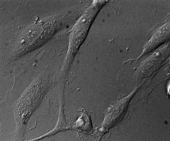
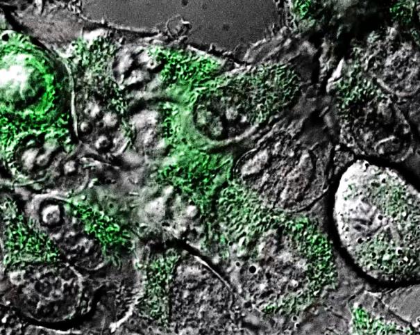
DIC imaging: Endothelia cell Mitochondria dynamics
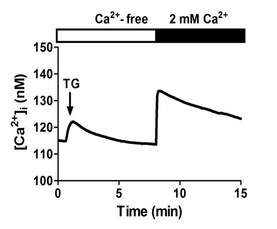
Ca2+ imaging: Thapsigargin (TG) stimulated Ca2+ jump
By Ming-Hsien Tsai from LSARC
FAQ/Contact:
Name:Chu, Chia-Ying
Tel No.:2371#23
E-mail:cychu@kmu.edu.tw